Histopathology
Submit biopsies, masses, surgical specimens, and other tissues for microscopic evaluation.
Submit tissue, biopsy, aspirate, fluid, and dermatology samples for pathology evaluation with clear handling requirements and case context.
Submit biopsies, masses, surgical specimens, and other tissues for microscopic evaluation.
Submit fine-needle aspirates, impression smears, and cellular samples for cytologic interpretation.
Submit body cavity fluids and other fluid samples for cellular evaluation and interpretation.
Submit skin biopsies and lesion samples when a dermatologic case needs tissue-level review.
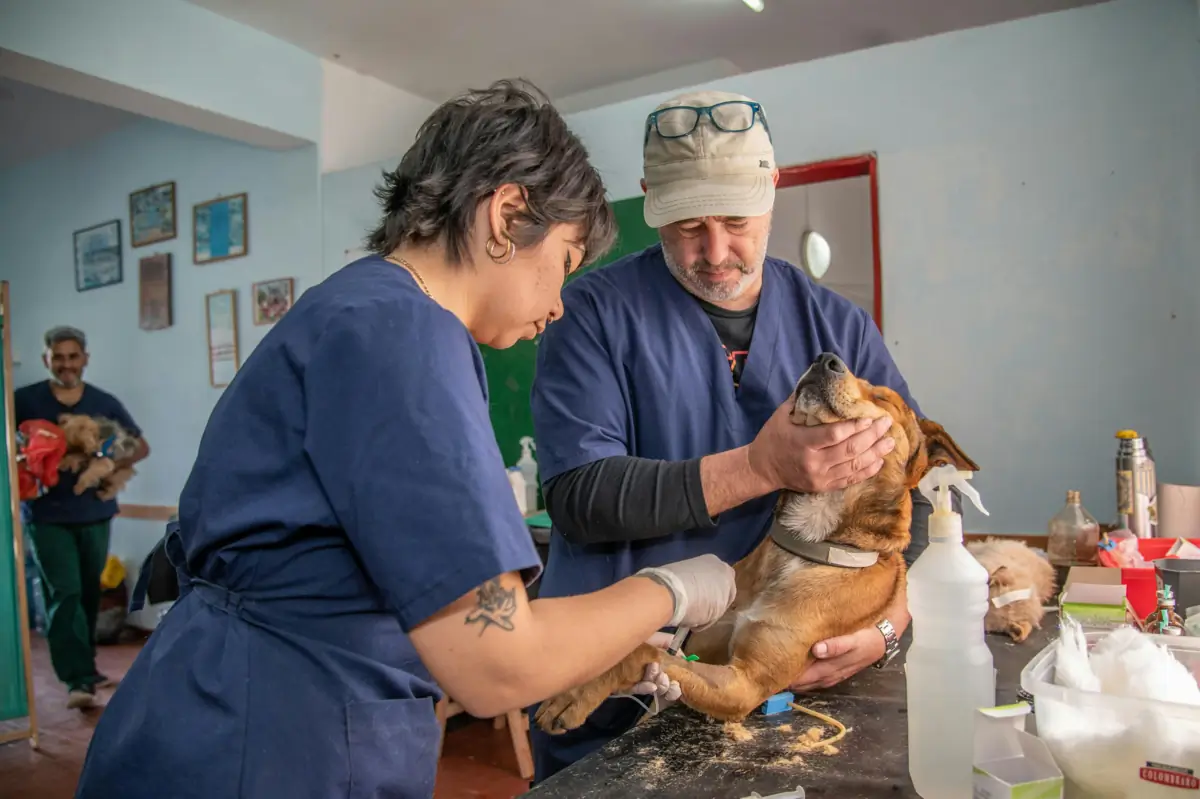
Pathology supports cases that require cellular or tissue-level interpretation beyond the physical exam, imaging, or routine laboratory findings.
A mass or lesion needs cytology or histopathology evaluation.
A biopsy is needed to characterize inflammation, infection, or neoplasia.
A removed mass needs tissue architecture and pathology interpretation.
A chronic or unusual skin lesion needs dermatopathology review.
An aspirate or fluid sample needs cellular interpretation.
Accurate labels, clinical context, and correct preservation help keep each finding connected to the right site and case.
Provide signalment, lesion duration, exact location, clinical findings, prior treatment, imaging findings, and relevant differentials when available.
Identify each container and slide by patient and anatomic site. When submitting multiple lesions, keep the labels matched to the submission form.
Place routine histopathology samples in 10% neutral buffered formalin unless B&L directs otherwise. Confirm container and volume before shipping.
Air-dry slides, label the sampled site, and keep them physically separated from formalin containers to protect staining quality.
Review B&L test options for cytology, fluid analysis, simple and complex histopathology, liver biopsy, and dermatopathology.
Use the B&L test menu to confirm the correct test code, specimen, fixative, container, handling requirements, and turnaround details before submission.
Search pathology tests| Test | Code | Turnaround |
|---|---|---|
| Cytology 1-2 days | 8000 | |
| Cytology - Fluid Analysis 1-3 Days | 8050 | |
| Dermatopathology 3-5 Days | 8020 | |
| Histopathology Complex, 1 Site 3-5 Days | 8011 | |
| View all 9 related tests | ||
Many practices save 15-30% a month on lab costs. Share your current lab setup, and B&L will review pricing, logistics, PIMS fit, contract timing, and next steps.
B&L accepts listed pathology submissions including tissue biopsies, masses, fine-needle aspirates, cytology slides, body fluids, liver biopsies, and dermatopathology samples. Confirm the exact test and specimen requirements before collection.
Cytology evaluates individual cells or small cell groups from aspirates, smears, or fluids. Histopathology evaluates preserved tissue architecture from a biopsy or surgical specimen.
Include patient identification, signalment, clinical history, exact sample site, collection method, lesion description, prior treatment, and relevant diagnostic findings.
They may be part of the same case, but slides should be packaged separately from formalin containers. Formalin vapor can interfere with cytology staining and slide quality.